Anatomy
The spinal cord is approximately the thickness of your little finger, and is made up of nerves (also called neurones). These nerves commence in the brain and run in a tunnelthrough the bones which make up the spine (the vertebra) in what is called the spinal canal.
The cord runs from the base of the skull all the way down into the pelvis.
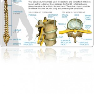
The spinal column is comprised of 33 separate bones called vertebrae: 7 cervical vertebrae (neck), 12 thoracic vertebrae (upper and middle back corresponding to the chest), 5 lumbar vertebrae (lower back), 5 sacral vertebrae (sacrum), and 4 fused coccygeal vertebrae (coccyx).
The nerves in the spinal cord carry both movement information TO muscles in the body and sensory information FROM the outside world back to the brain. These are called central nerves.
They are unique in that they cannot repair themselves if damaged or cut: they cease to function, and so cannot carry messages. The areas of the body to which they correspond also cease to function properly: no sensations are relayed from the outlying regions to the brain and no movement messages can be sent to the muscles of the limbs, trunk and other body parts.
Exiting from the spinal cord and running to the trunk and limbs are peripheral nerves, which are different in that they can repair themselves or be surgically repaired if they are damaged.
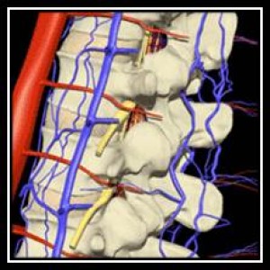
Like other parts of the body, the nerves in the central and peripheral nervous system require blood carrying oxygen to provide energy to the nerve cells themselves. This oxygen arrives via arteries, and is drained away by veins from all areas of the spine.
Damage to the spinal cord itself, whether it is severed, crushed, compressed or bent can cause disruption to the electrical impulses which carry information to and from the brain in the nerves, and a spinal cord injury results.
Similarly, swelling around the cord pressing upon it, disruption to the blood flow in the arteries coming to the spinal cord or restriction of outflow via the veins draining the spinal cord can also result in spinal cord injury.

The spinal cord runs in a bony tunnel within the vertebra of the spine.
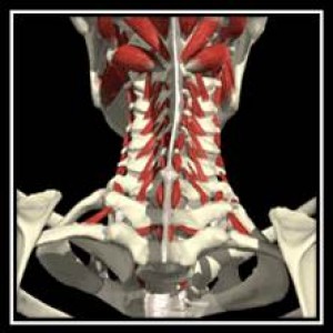
These vertebra are separated by an intervertebral disc of a shock-absorbing, elastic cartilage, and surrounded by muscles which help move the bones and thus turn the associated part of the body, such as the neck, chest and the lower back.
The bones in the neck (the cervical vertebrae) are most susceptible to injury because they do not have the protection afforded by the ribs in the trunk or the organs within the abdomen. They are thus able to move to a greater extent forward, backwards and sideways, as well as rotating from left to right.
All vertebra are protected from excessive movement by the individual architecture of each bone, and by a series of ligaments, tendons and muscles which prevent excessive movement of one vertebra on another are disrupted. These structures act as springs to both initiate and also to limit the full range of movement.


